What determines whether a blood vessel remains elastic for decades or ruptures unexpectedly? It is a question that not only keeps doctors, but especially patients awake. Within the CELLSYSTEMICS project, Koen Reesink of the Cardiovascular Research Institute Maastricht (Maastricht UMC) investigates how the human vascular wall works, reacts and sometimes fails, using a unique human measurement model.
CELLSYSTEMICS is a research platform that studies the behavior of human vascular cells under conditions that occur in the body. The focus is on the smooth muscle cells in the walls of large blood vessels. These cells do not contract spontaneously, like heart muscle cells, but are continuously stretched by blood pressure. “With every heartbeat, the artery expands slightly,” explains project leader Koen Reesink. “That is necessary to relieve the heart. That stretching is not a side issue, it is an important signal for the cell: keep doing what you are doing.” Yet, he says, that mechanical behavior is hardly tested in cell biology. “We mainly look at biochemical markers to determine whether a cell is behaving ‘healthily’. But we still know little about what that cell actually does when it is under pressure.”
The project not only looks at individual cells, but also at their behavior as a group. As part of living, deformable tissue. “Cells sense each other, communicate, respond to each other’s behavior,” says Reesink. “If one cell starts doing something different, the others notice that too.” Yet this mutual interplay is hardly ever included in experiments. According to Reesink, this is a major shortcoming in much biomedical research. “Classical experimental biology tries to work towards a homogeneous population of cells or people. While in reality, diversity is the norm.”
CELLSYSTEMICS in three workflows
Workflow 1 – Stem cell differentiation
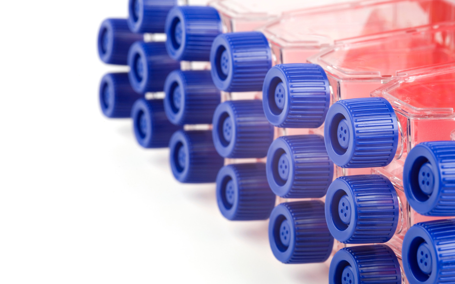
Stem cells, collected from patient blood, are converted into smooth muscle cells of the vascular wall using specific media and protocols.
Workflow 2 – Cellular level measurements
Micro-indenting and fluorescence microscopy are used to measure how individual cells respond to mechanical loading: for example in calcium response and cytoskeletal dynamics.
Workflow 3 – Functional tissue model
Cells are brought together in a bioreactor that simulates real vessel wall loading. This allows us to look at force generation, response to drugs and the behaviour of cells as in a network.
Ticking time bomb
Patients with an aneurysm – an enlargement of the artery – often feel like a ticking time bomb, says Reesink. “They know that things can go wrong, but no one knows when. That creates enormous uncertainty. They want to understand what is happening.” CELLSYSTEMICS therefore not only investigates what goes wrong, but also what functions normally. “Why doesn’t everyone get an aneurysm? That is the real question – and if we understand it, we can provide better answers.”
In the long term, the project can contribute to patient-specific predictions, although that remains a pipe dream for now. “We may one day be able to deduce from a blood sample what the course of the disease looks like,” says Reesink. Testing treatments is also part of the project: in cultured vascular tissue, it is being examined whether medicines can influence the 'attitude' of cell groups. At the moment, growing patient-own cells still takes months, but that process must become faster. “If we move towards a month, clinical application will come closer.”
Beating heart muscle cells
Although CELLSYSTEMICS focuses on the vascular wall, its scope is broader. Functioning heart muscle cells have now also been grown in the lab — beating and all. But even there, the behavior of cells under load is still difficult to measure. “The mechanical environment determines how cells behave,” says Reesink. “Stretch, pressure, adhesion — all those forces give signals, and they are often local.” The platform is therefore relevant for any type of tissue that is subject to physical stress: from heart and lung tissue to muscles or even tumor cells.
Mutual trust
Collaboration is essential for the success of CELLSYSTEMICS, Reesink emphasizes. “You have to learn to speak each other’s language — and dare to say when you don’t understand each other. Only then will you make progress.” This applies to collaboration between disciplines, but also with companies. “You simply need their expertise, otherwise you won’t get there.” According to Reesink, it’s all about mutual trust. “I have given partners confidence that I would make a whole out of their knowledge. Without that synergy, I would never have been able to continue this project.”
CELLSYSTEMICS is an initiative of the departments of Biomedical Engineering, Cardiothoracic Surgery and Biochemistry of the Cardiovascular Research Institute Maastricht, and is made possible by the Public-Private Partnership subsidy made available by Health-Holland – Top-Sector Life Sciences & Health, Samenwerkende GezondheidsFondsen (SGF) and ZonMW.
Want to know more? Koen Reesink is one of the speakers at the Life Science Live event on Tuesday, May 19, in the Plus Ultra Building on the Utrecht Science Park Campus. View the program and register at the website.